The third eyelid in Golden Retrievers is caused by five main conditions: cherry eye, Horner’s syndrome, keratoconjunctivitis sicca, conjunctivitis, and systemic illness. A third eyelid that stays visible beyond a few minutes after waking up is not normal. It means the eye needs assessment, and some causes behind it require same-day veterinary contact.
In my practice, dog third eyelid showing is one of the presentations that owners most frequently misread as harmless. Goldens are prone to cherry eye due to weak connective tissue attachment of the nictitating gland, and they also develop KCS at elevated rates. Both conditions cause the third eyelid to protrude, but they need completely different treatment paths.
According to the Golden Retriever Lifetime Study, conducted by the Morris Animal Foundation, Golden Retrievers have a documented elevated risk for immune-mediated ocular conditions. These include keratoconjunctivitis sicca, a leading cause of dog third eyelid, showing that most general guides fail to connect to third eyelid protrusion in this breed.
Contents
- 1 Dog Third Eyelid Showing: The 5 Causes That Appear in Golden Retrievers
- 2 Why Golden Retrievers Show the Third Eyelid More Than Other Retrievers
- 3 Dog Third Eyelid Showing in Golden Retrievers by Age: Puppy, Adult, and Senior
- 4 What Most Dog Third Eyelid Guides Get Wrong About Golden Retrievers.
- 5 The GRI Third Eyelid Check Protocol: What to Look For Before You Call.
- 6 When to Call the Vet: Urgent vs. Monitor Table.
- 7 EXPERT INSIGHT CALLOUT.
- 7.1 What causes dog third eyelid to show in Golden Retrievers?
- 7.2 Is dog third eyelid showing always a sign of illness?
- 7.3 What does dog third eyelid showing and squinting mean together?
- 7.4 Why do dogs eyelids sometimes roll inward and cause problems?
- 7.5 What does a red dog eyeball alongside a visible third eyelid mean?
- 7.6 What is cherry eye in dogs, and how is it treated?
- 7.7 Can a dog third eyelid showing go away on its own?
- 7.8 What is Horner’s syndrome in dogs?
- 7.9 How do vets treat a prolapsed third eyelid in dogs?
- 7.10 What happens if a cherry eye in a dog goes untreated?
- 7.11 Do Golden Retrievers get cherry eye more than other breeds?
- 7.12 How does Horner’s syndrome affect Golden Retrievers differently from Labradors?
- 7.13 What does dog third eyelid showing alongside KCS mean for a Golden Retriever?
- 7.14 At what age do Golden Retrievers typically develop cherry eye?
- 7.15 My Golden Retriever has a red lump in the corner of one eye. What do I do right now?
- 8 Conclusion.
Dog Third Eyelid Showing: The 5 Causes That Appear in Golden Retrievers
Dog eye problems have a lot of conditions; among them, the dog’s third eyelid always has a different cause. In Golden Retrievers, five conditions account for the vast majority of presentations I see.
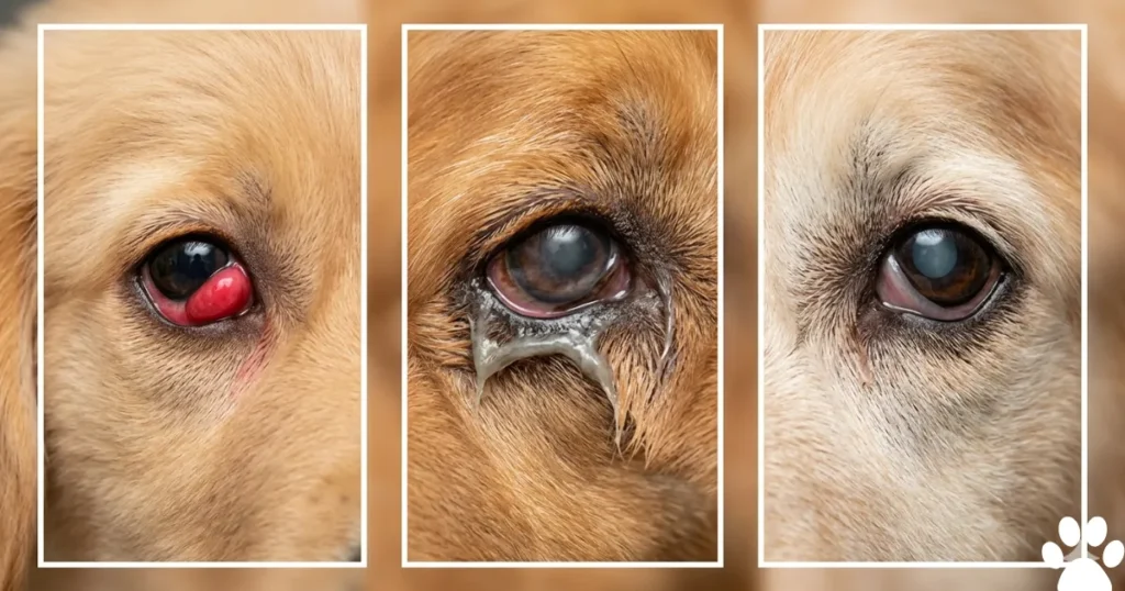
Cherry Eye
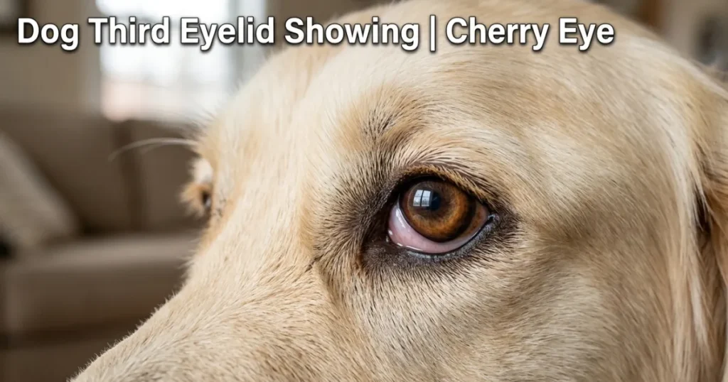
Cherry eye is the prolapse of the nictitans gland, the tear-producing gland attached to the base of the third eyelid. When the connective tissue holding it in place weakens or tears, the gland flips forward and appears as a red, fleshy, round mass in the inner corner of the eye. Dog third eyelid showing from cherry eye is usually obvious. The red mass is visible and distinct from the surrounding tissue.
Golden Retrievers develop cherry eye more frequently than Labradors because they have weaker ligament attachments at the nictitans gland base. The AVMA recognizes certain breeds, including Golden Retrievers, as predisposed to this condition. Left untreated, cherry eye reduces tear production and leads to secondary KCS.
Horner’s Syndrome
Horner’s syndrome is a neurological condition caused by damage or disruption to the sympathetic nerve pathway supplying the eye. It produces four classic signs together: the third eyelid rising up across the dog eyeball, the upper eyelid drooping, the lower eyelid rising slightly, and the pupil becoming smaller than the other side. The eye looks sunken.
This is the cause I worry about most when owners describe dog third eyelid showing alongside unequal pupils. Horner’s syndrome can signal a neck injury, a middle ear problem, or a chest mass pressing on the nerve pathway. It needs urgent investigation.
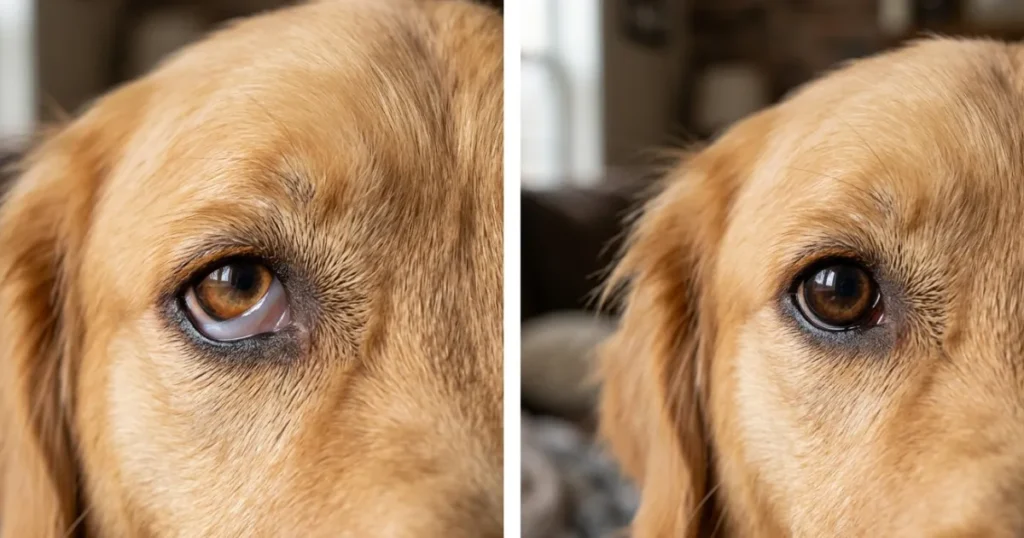
Keratoconjunctivitis Sicca
KCS causes dog third eyelid to show because chronic corneal dryness triggers the nictitating membrane to slide partially across the dog eyeball surface as a protective reflex. The third eyelid sits higher than normal, and the eye looks dull. Owners also notice thick, grey-white ropy discharge alongside the protrusion.
KCS is immune-mediated in the majority of Golden Retriever cases. The AAHA recognizes immune-mediated KCS as a condition of elevated concern in Golden Retrievers compared to the general dog population.
Conjunctivitis
Bacterial or viral conjunctivitis causes eye irritation and swelling of the conjunctival tissue. Swollen tissue pushes the third eyelid forward passively. Dog third eyelid showing from conjunctivitis appears alongside red, swollen conjunctiva and either watery or yellow-green discharge. The eyelid itself still moves normally.
Systemic Illness
Severe dehydration, fever, extreme pain, or wasting illness can cause a bilateral dog third eyelid showing because the fat pad behind the dog eyeball shrinks, allowing the eyeball to sink back slightly into the socket. The third eyelid then covers more of the eye surface than usual. If both third eyelids are showing at the same time and your golden seems unwell, contact your vet the same day.
Why Golden Retrievers Show the Third Eyelid More Than Other Retrievers
I’ll be direct: Golden Retrievers are not the same as Labradors when it comes to third eyelid conditions. Three factors separate them.
Connective Tissue Laxity
Golden Retrievers have looser connective tissue at the nictitans gland attachment than most retrievers. This is the structural reason cherry eye appears more often in Goldens. The gland prolapse that causes dog third eyelid to show can happen in a single vigorous head shake in a puppy with already loose ligaments. I’ve seen it happen during routine play in dogs as young as 8 weeks.
In my practice, Goldens account for a disproportionate share of cherry eye presentations compared to other retriever breeds I see. This matters because once the gland prolapses, surgical repositioning rather than removal is the standard of care. Removing the gland reduces tear production by approximately 30%, which compounds the KCS risk Goldens already carry.
The KCS Immune Connection
Golden Retrievers develop immune-mediated KCS at higher rates than Labradors. The same systemic immune dysregulation documented in the Golden Retriever Lifetime Study population that increases cancer risk also affects lacrimal gland tissue. When KCS develops, the corneal surface loses adequate lubrication, and the third eyelid begins sliding across the dog eyeball as a compensatory reflex.
What I tell owners: If the third eyelid is showing and the discharge is grey-white and ropy rather than watery, think KCS before conjunctivitis. Because in a Golden over age 4, KCS is the higher-probability cause of that presentation.
The Nerve Pathway Factor
Golden Retrievers are physically active dogs with large necks and strong musculature. Neck injuries from pulling hard on a lead, jumping from heights, or rough play can compress or stretch the sympathetic nerve pathway that runs through the neck toward the eye. That compression causes Horner’s syndrome, and dog third eyelid is showing. I’ve seen Horner’s syndrome triggered by a single aggressive leash pull in a young male Golden who had no prior eye health history. For a general health guide, visit our general health blogs.
Dog Third Eyelid Showing in Golden Retrievers by Age: Puppy, Adult, and Senior
Golden Retriever Puppies (8 Weeks to 18 Months).
Cherry eye is the dominant cause of dog third eyelid showing in Golden Retriever puppies. The connective tissue is at its weakest in the first 12 months of life. Most cherry eye in Goldens appears before 18 months of age.
The most common mistake I see is owners waiting two to three weeks, hoping it will self-correct. It’s understandable because the puppy often acts completely normal, but in Goldens specifically, waiting causes the prolapsed gland to swell further and makes surgical repositioning harder. Seek a vet exam within 48 hours of first noticing it.
Dogs eyelids in puppies can also show entropion, an inward rolling of the lid margin, which produces a dog third eyelid showing a response from corneal irritation. Entropion sometimes self-corrects by 16 weeks. If it persists beyond that point, surgical correction is needed.
Adult Golden Retrievers (2 to 7 Years).
Dog third eyelid of a dog in adult Goldens most commonly indicates KCS or conjunctivitis. When a Golden owner calls me about this concern in a dog between the ages of 4 and 6, the first question I ask is whether the eye looks dull or dry compared to the other side. A dull, slightly sunken appearance with ropy discharge tells me KCS is more likely than infection.
Horner’s syndrome also presents in this age window, often following a physical incident. Any adult Golden Retriever showing dog third eyelid alongside unequal pupils needs same-day veterinary attention.
Senior Golden Retrievers (8 Years and Older).
Senior Goldens face three overlapping causes of dog third eyelid showing. KCS progresses with age as immune-mediated lacrimal gland damage accumulates. Systemic illness becomes more likely, and the fat pad behind the dog eyeball naturally thins with age and body condition loss, causing the eye to sit slightly deeper in the socket.
Pigmentary uveitis, a Golden Retriever-specific inflammatory condition recognized by the GRCA, can also produce secondary third eyelid irritation and protrusion in senior dogs. Any senior Golden showing dog third eyelid alongside iris darkening or cysts at the pupil margin needs an intraocular pressure check at the same visit.
What Most Dog Third Eyelid Guides Get Wrong About Golden Retrievers.
Most dog third eyelid guides treat cherry eye as the default explanation for every case. For Golden Retrievers, this misses two clinically distinct and more serious causes.
Here’s the problem. Cherry eye produces a visible red, fleshy mass in the eye corner. It is hard to miss. Horner’s syndrome produces a third eyelid that rises up quietly, with no mass, no redness in the corner, and no discharge. The eye just looks different, with a smaller pupil and a slightly drooping upper lid. Owners see dog third eyelid showing and assume cherry eye because that’s what every guide describes first. They wait for a lump that never appears. Meanwhile, the underlying nerve compression that caused the Horner’s syndrome goes uninvestigated.
The second missed cause is KCS. KCS is immune-mediated and chronic. It produces dog third eyelid, showing because the corneal surface becomes so dry that the nictitating membrane compensates by sliding across the dog eyeball. Treating this presentation with antibiotic drops, the standard recommendation in most guides, does nothing for KCS and allows corneal scarring to progress.
In April 2025, a 5 years old male Golden presented with dog third eyelid in the right eye. The owner had been applying over-the-counter lubricating drops for two weeks after reading that third eyelid protrusion usually resolves on its own. The right pupil was visibly smaller than the left. The upper lid had mild ptosis. Neurological assessment confirmed Horner’s syndrome. Imaging identified a mild cervical disc irregularity at C3 to C4 consistent with a nerve compression event. The owner’s takeaway: unequal pupils alongside dog third eyelid showing are never a wait and see presentation.
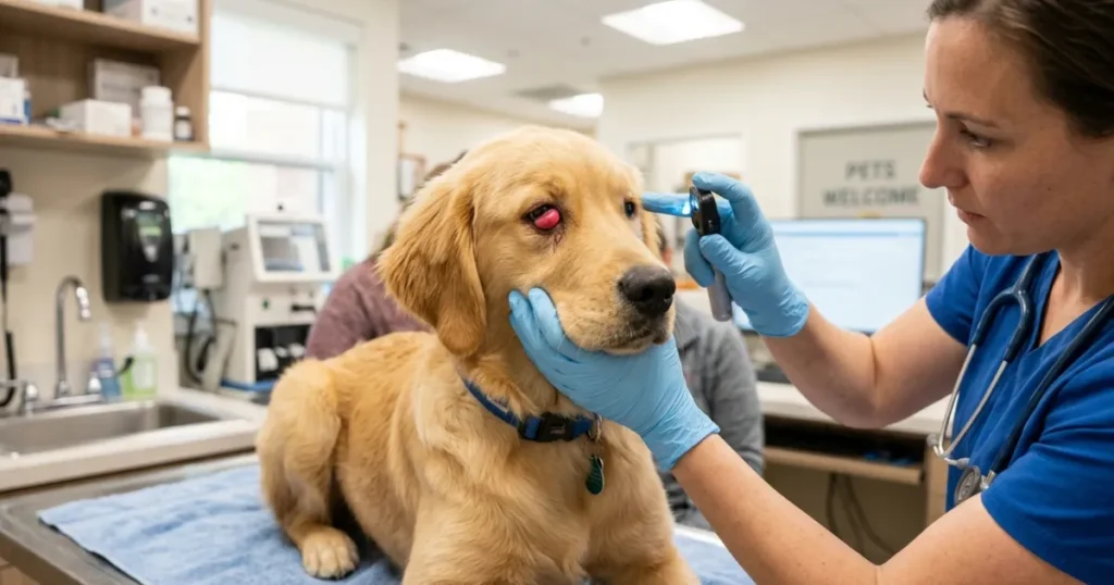
The GRI Third Eyelid Check Protocol: What to Look For Before You Call.
This is the three-step framework I walk Golden owners through when they describe dog third eyelid showing over the phone. Complete all three steps before you call, so you can describe exactly what you see.
STEP 1—Check the Appearance of the Third Eyelid.
Look at the inner corner of the affected eye in good light.
If you see a red, fleshy, round mass, this is likely a cherry eye. Call your vet today for an appointment within 48 hours.
If the third eyelid is simply elevated with no mass, no redness in the corner, and no discharge, move to Step 2.
If the third eyelid is elevated and the eye looks dull with grey-white ropy discharge, it is likely KCS. Call your vet today.
STEP 2—Check Both Pupils at the Same Time.
Stand in front of your Golden in normal indoor lighting. Cover one eye gently and look at the uncovered pupil size. Then swap and check the other. Both pupils should be the same size.
If the pupils are unequal, this is a Horner’s syndrome signal until your vet rules it out. Call your vet the same day.
If the pupils are equal, move to Step 3.
STEP 3—Check Duration and Paired Behavior.
If the dog third eyelid showing appears right after waking and clears within 10 minutes: monitor for 24 hours. This can be a normal sleep-related response.
If the dog third eyelid showing has been present for more than one hour while your Golden is awake and active, call your vet today.
If your Golden is squinting, pawing at the eye, or avoiding light alongside dog third eyelid showing: Call your vet immediately. Read more about squinting paired with third eyelid changes in our dog squinting guide.
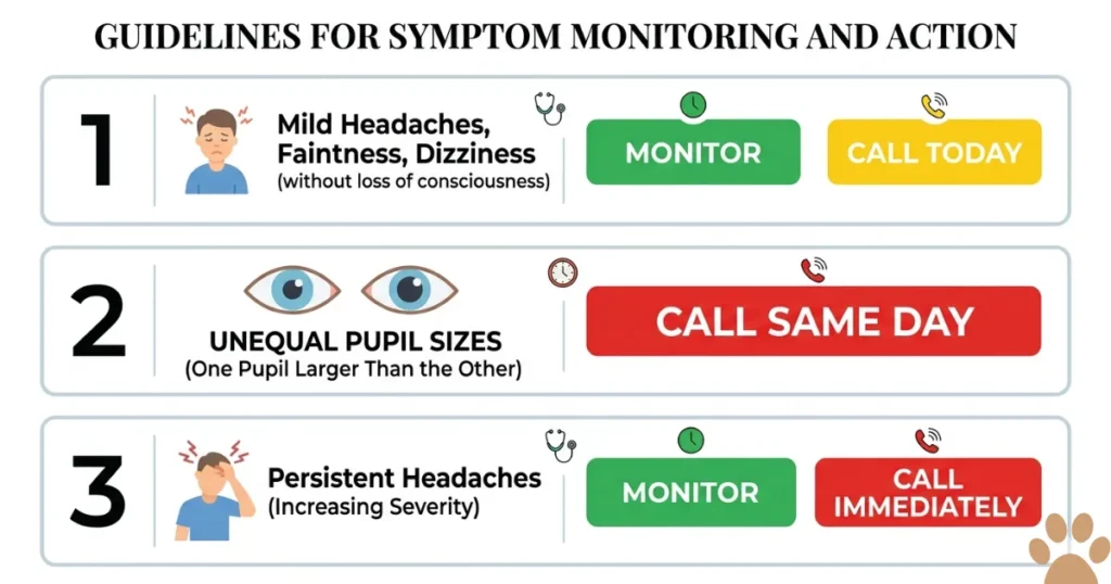
When to Call the Vet: Urgent vs. Monitor Table.
| CALL VET IMMEDIATELY 🔴 | MONITOR AT HOME 24 HRS 🟡 |
| Red fleshy mass visible in eye corner (cherry eye) | The third eyelid visible after sleep and clears within 10 minutes |
| Unequal pupils alongside third eyelid protrusion | Both eyes look symmetrical |
| Drooping upper eyelid on the affected side | Your Golden is eating, drinking, and acting normally |
| Dog eyeball looks sunken or asymmetric | Mild redness with no discharge and no squinting |
| Bilateral third eyelid showing with lethargy or fever | — |
| Third eyelid showing alongside heavy squinting | — |
| Grey-white ropy discharge with a dull corneal surface | — |
If monitoring and any URGENT sign develop within 24 hours, stop monitoring. Call your vet immediately.
EXPERT INSIGHT CALLOUT.
“The cases that concern me most are the ones where the owner says the third eyelid has been showing for two weeks, and the dog seems fine otherwise. In Golden Retrievers, that two-week window is exactly when Horner’s syndrome investigations should have started and when a KCS diagnosis would have saved the cornea from early scarring. Dog third eyelid showing that it lasts more than 24 hours in a Golden always earns a vet visit. That’s the threshold I use with every owner I talk to.”
What causes dog third eyelid to show in Golden Retrievers?
Dog third eyelid in Golden Retrievers is caused by cherry eye, Horner’s syndrome, keratoconjunctivitis sicca, conjunctivitis, and systemic illness. Golden Retrievers develop cherry eye and KCS at higher rates than Labradors due to connective tissue laxity and their immune profile, per AVMA and AAHA breed data.
Is dog third eyelid showing always a sign of illness?
Dog third eyelid showing immediately after sleep can be normal and clears within 10 minutes. A third eyelid that stays visible while your dog is fully awake and active for more than one hour is not normal. It signals one of five conditions that need veterinary assessment, not home treatment.
What does dog third eyelid showing and squinting mean together?
Dog third eyelid showing and squinting together indicates ocular pain alongside the protrusion. In Golden Retrievers, this combination most commonly points to a corneal ulcer secondary to KCS or cherry eye or to acute uveitis. Both are same-day vet presentations. Do not apply drops before the exam.
Why do dogs eyelids sometimes roll inward and cause problems?
Dogs eyelids roll inward due to entropion, a condition where the lid margin turns toward the eye surface. Entropion causes lashes to rub the cornea with every blink, producing pain, tearing, and secondary third eyelid elevation. Golden Retrievers are among the breeds with documented entropion predisposition, per AVMA breed data.
What does a red dog eyeball alongside a visible third eyelid mean?
A red dog eyeball alongside a visible third eyelid suggests active inflammation inside the eye or on the surface. In Golden Retrievers, this combination often indicates canine glaucoma, acute uveitis, or severe conjunctivitis with secondary protrusion. All three are same-day vet presentations. A red dog eyeball with a fixed pupil is an emergency.
What is cherry eye in dogs, and how is it treated?
Cherry eye in dogs is the prolapse of the nictitans gland, the tear-producing gland at the base of the third eyelid. Treatment is the surgical repositioning of the gland back into place. Removal is no longer the standard of care because it reduces tear production by approximately 30%, increasing KCS risk in already predisposed breeds like Golden Retrievers.
Can a dog third eyelid showing go away on its own?
Dog third eyelid, showing from mild irritation, can resolve within a few hours. Cherry eye does not self-correct and requires surgery. Horner’s syndrome requires investigation for the underlying cause. KCS requires lifelong prescription eye drops. Any dog third eyelid showing lasting more than 24 hours in a Golden needs a vet assessment.
What is Horner’s syndrome in dogs?
Horner’s syndrome in dogs is a neurological condition caused by disruption to the sympathetic nerve supply to the eye. It produces four signs together: third eyelid elevation, upper eyelid drooping, lower eyelid rising, and a smaller pupil on the affected side. Causes include neck injury, middle ear disease, and chest masses compressing the nerve pathway.
How do vets treat a prolapsed third eyelid in dogs?
Vets treat a prolapsed third eyelid, or cherry eye, with surgical repositioning using a pocket technique or imbrication suture to secure the gland back in its normal position. Anti-inflammatory drops manage post-surgical swelling. Golden Retrievers may need KCS monitoring after surgery because the gland is critical for their tear film.
What happens if a cherry eye in a dog goes untreated?
Untreated cherry eye causes the prolapsed nictitating membrane gland to swell progressively from chronic exposure. Tear production declines as the gland is damaged. Secondary KCS develops, leading to chronic dry eye, corneal ulceration, and scarring. In Golden Retrievers, who already carry elevated KCS risk, untreated cherry eye accelerates this progression significantly.
Do Golden Retrievers get cherry eye more than other breeds?
Yes. Golden Retrievers develop cherry eye at higher rates than Labradors and most other retrievers because of weaker connective tissue attachment at the nictitating membrane. Do Golden Retrievers get cherry eye more than other breeds? gland base. The AVMA recognizes Golden Retrievers as among the predisposed breeds. Most cherry eye in Goldens appears before 18 months of age, though it can occur in adults.
How does Horner’s syndrome affect Golden Retrievers differently from Labradors?
Horner’s syndrome in Golden Retrievers is more frequently triggered by neck and cervical spine events because Goldens are more prone to pulling hard on a lead and jumping from heights during play. The sympathetic nerve pathway through the neck is vulnerable to compression in these incidents. Labradors show a similar mechanism, but Golden Retrievers present with cervical-origin Horner’s more often in my clinical experience.
What does dog third eyelid showing alongside KCS mean for a Golden Retriever?
Dog third eyelid, showing alongside KCS in a Golden Retriever, means the corneal surface is so dry that the nictitating membrane is compensating by sliding partially across the eye. According to the AAHA, Golden Retrievers develop immune-mediated KCS at elevated rates. Treatment with cyclosporine or tacrolimus drops addresses the immune cause. Antibiotic drops alone do not help.
At what age do Golden Retrievers typically develop cherry eye?
Golden Retrievers typically develop cherry eye between 8 weeks and 18 months of age, when the connective tissue attachment of the nictitating gland is at its weakest. Later-onset cherry eye in adult Goldens can occur but it is less common. Any dog third eyelid showing a red mass in the eye corner before age 2 should be examined within 48 hours.
My Golden Retriever has a red lump in the corner of one eye. What do I do right now?
Call your vet today and request an appointment within 48 hours. A red lump in the inner corner of a Golden Retriever’s eye is cherry eye until proven otherwise. Do not wait for it to self-correct. Do not try to push it back manually. Early surgical repositioning has a higher success rate than surgery performed after the gland has been exposed and swollen for weeks.
Conclusion.
Dog third eyelid showing in Golden Retrievers is never a dismiss-and-wait situation beyond 24 hours. Five conditions cause it: cherry eye, Horner’s syndrome, KCS, conjunctivitis, and systemic illness. Goldens are predisposed to cherry eye and KCS at rates that make them more vulnerable than most retrievers, and Horner’s syndrome presentations in this breed are frequently missed because owners wait for a symptom that looks like cherry eye but never appears.
Use the GRI Third Eyelid Check Protocol before you call your vet. Check the appearance of the third eyelid, check both pupils for symmetry, and then check how long the protrusion has been present. Those three steps give your vet a precise picture before you arrive and help them triage your call correctly. Has your Golden ever had a dog third eyelid showing episode that turned out to be more serious than you expected? Tell me what age your dog was, what the first sign looked like, and how long it took to get a diagnosis. Real accounts from Golden owners help others in this community catch the same signs earlier and act faster.

Dr. Nabeel A.
Dr. Nabeel Akram is a Doctor of Veterinary Medicine with more than five years of hands-on experience in animal health, canine nutrition, and preventive care. He is a registered veterinarian with the Pakistan Veterinary Medical Council (PVMC), the statutory body regulating veterinary practice in Pakistan. As the founder of Golden Retriever Insight, Dr. Akram writes and medically reviews every health, nutrition, and grooming guide published on the site. His clinical interests include canine oncology, epilepsy management, and breed-specific nutrition for large breeds — the core topics this site covers. Every article is checked against current veterinary literature and sources such as the Merck Veterinary Manual, AVMA guidance, and peer-reviewed research.