
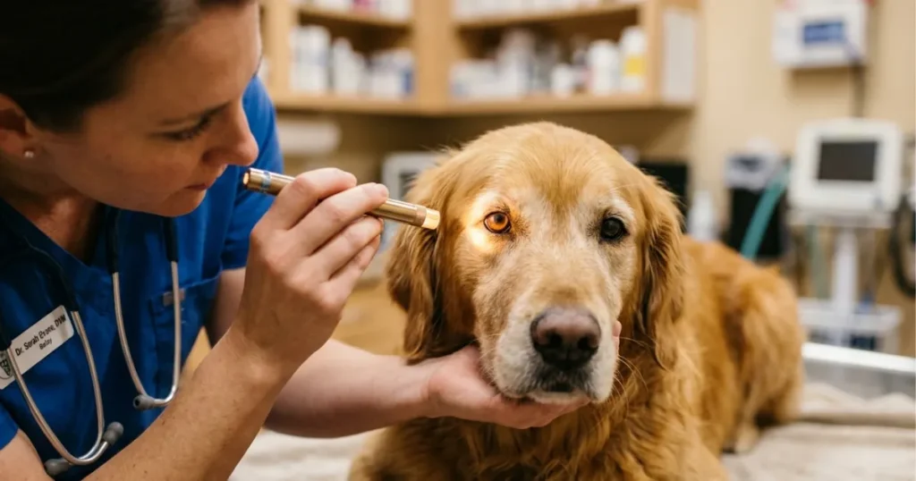
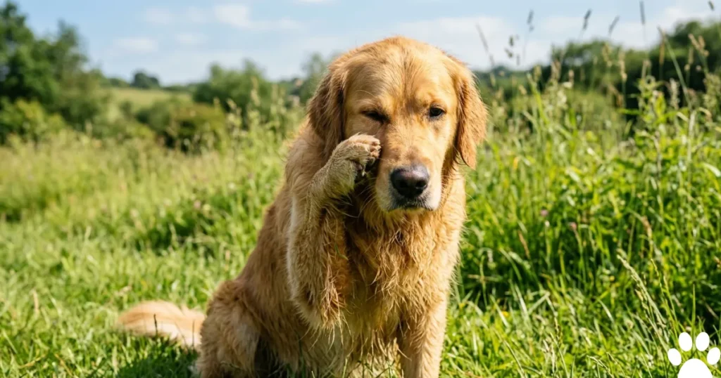
Dog eye health issues in Golden Retrievers fall into six visually distinct categories. The appearance of the discharge, the eye surface, and the iris color tells you which category you’re dealing with before your vet runs a single test. Most online picture guides show generic red eyes. That’s not enough to act on.
In my practice, the owners who catch problems early are the ones who know specifically what to look for. Not just “redness.” Whether the redness sits in the white of the eye, the inner eyelid, or around the cornea. Not just “discharge.” Whether it’s watery, ropy, and grey-white, or yellow-green and crusting the eyelid shut overnight.
According to the Golden Retriever Lifetime Study, conducted by the Morris Animal Foundation, Golden Retrievers carry a documented elevated lifetime risk for heritable ocular conditions, including pigmentary uveitis and progressive retinal atrophy. Both conditions have specific, identifiable visual presentations that differ meaningfully from generic canine conjunctivitis.
Contents
- 1 Dog Eye Health Issues: What Each Visual Sign Actually Means in Golden Retrievers.
- 2 Why Golden Retrievers Show Dog Eye Health Issues Differently Than Other Breeds.
- 3 Dog Eye Health Issues in Golden Retrievers by Age: Puppy, Adult, and Senior.
- 4 What Most Dog Eye Infection Picture Guides Get Wrong About Golden Retrievers?
- 5 The GRI Visual Symptom Classifier: Matching What You See to What It Means.
- 5.1 SIGN 1—Clear, watery, both eyes open, no crust, no squinting.
- 5.2 SIGN 2—Thick, ropy, grey-white discharge with a dull corneal surface.
- 5.3 SIGN 3—Yellow-green discharge crusting the eyelid, unilateral first.
- 5.4 SIGN 4—Iris darkening, brownish color, and small cysts at the pupil margin.
- 5.5 SIGN 5—Dense white lens opacity with no tapetal glow under a flashlight.
- 5.6 SIGN 6—Bulging eye, asymmetric pupils, one eye visibly larger.
- 6 When to Call the Vet: Urgent vs. Monitor Table.
- 7 EXPERT INSIGHT CALLOUT.
- 7.1 What dog eye health issues should owners watch for?
- 7.2 How do I identify dog eye health issues at home?
- 7.3 What do dog eye infections look like in pictures?
- 7.4 How do dog eye problems look in pictures vs. real life?
- 7.5 What are the common eye ailments in dogs by appearance?
- 7.6 What is the best eye care routine for dogs?
- 7.7 Can a dog eye infection spread to humans?
- 7.8 What does normal dog eye discharge look like?
- 7.9 How do vets diagnose eye problems in dogs?
- 7.10 What happens if a dog eye infection goes untreated?
- 7.11 Do Golden Retrievers get more eye infections than other breeds?
- 7.12 What does pigmentary uveitis look like in a Golden Retriever?
- 7.13 How do Golden Retrievers show signs of dry eye differently from Labradors?
- 7.14 At what age do Golden Retrievers show signs of progressive retinal atrophy?
- 7.15 My Golden has green discharge and won’t open one eye—is this an emergency?
- 8 Conclusion.
Dog Eye Health Issues: What Each Visual Sign Actually Means in Golden Retrievers.
Six visual signs account for nearly every golden retriever I see. Each point corresponds to a different condition, and each requires a different response. Here are the details of the dog’s eye problems.
Clear, Watery Discharge from One or Both Eyes.
Watery discharge with no crust, no color, and no pain behavior is the lowest-urgency presentation. In Goldens, it usually signals environmental irritation from pollen, grass seed, or wind exposure. It can also reflect early allergic conjunctivitis.
Here’s what makes this golden-specific. Goldens with longer facial coats have periocular hair that contacts the corneal surface repeatedly. That repeated contact produces low-grade tearing that looks like discharge but is actually a protective reflex. If you see clear tearing only after outdoor time that clears within an hour indoors, it’s almost always mechanical irritation rather than infection.
Monitor for 24 hours. If it persists or adds any color, call your vet.
Thick, Ropy, Grey-White Mucoid Discharge.
This is the sign most owners get wrong. Thick, stringy, grey-white discharge collecting at the inner corner that drags when wiped is keratoconjunctivitis sicca, not a bacterial infection. The two look similar in pictures. They are not in the same condition, and they don’t respond to the same treatment.
KCS discharge is grey-white and ropy because the aqueous layer of the tear film is deficient. The mucin component concentrates without the fluid to thin it. Bacterial discharge is yellow-green and tends to crust the eyelid shut overnight. If your Golden’s eye looks like it’s producing paste instead of liquid, that’s a KCS sign.
I’ve seen three cases this year where owners bought over-the-counter antibiotic drops for what was actually KCS. The drops did nothing. The cornea worsened because the underlying tear deficiency continued untreated. KCS in Goldens requires cyclosporine or tacrolimus drops, both available only by prescription.
Yellow-Green Mucopurulent Discharge.
Yellow or green discharge—especially when it crusts the eyelid shut overnight—indicates active bacterial involvement. In Goldens, this most commonly follows a viral episode or develops secondary to a corneal injury.
The key detail: it’s almost always unilateral first. If both eyes produce yellow-green discharge simultaneously, suspect a systemic cause. Both require immediate vet contact.
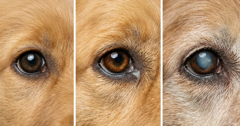
Iris Darkening or Cysts at the Pupil Margin.
This is the visual sign that no picture guide covers adequately. Pigmentary uveitis in Golden Retrievers presents as progressive iris darkening. The iris looks muddier, browner, or less golden than it used to. In some cases, small fluid-filled cysts are visible at the edge of the pupil, appearing as tiny dark bubbles.
The GRCA officially recognizes pigmentary uveitis as a Golden Retriever-specific condition, typically appearing in dogs aged 7 to 10 years. Owners frequently dismiss iris darkening as cosmetic aging. It’s not. Left unmanaged, it leads to secondary canine glaucoma and irreversible cataract formation.
Dense White or Grey Lens Opacity.
A white or grey opacity over the lens indicates either a cataract or nuclear sclerosis. The practical test: shine a light toward the eye. Nuclear sclerosis still shows the tapetal glow through the haze. A cataract blocks it. No glow and a dense white lens mean scheduling a vet exam promptly.
Bulging, Asymmetric, or Fixed Eye.
A visibly enlarged or asymmetric eye, a pupil that doesn’t respond to light, or an eye that appears to protrude more than usual are signs of canine glaucoma until proven otherwise. Intraocular pressure above 30 mmHg damages the optic nerve within hours in dogs. Don’t search for pictures and compare. Call your vet or an emergency animal hospital immediately.
Why Golden Retrievers Show Dog Eye Health Issues Differently Than Other Breeds.
The following are the details of dog eye health issues:
The Coat and Anatomy Factor.
Golden Retrievers have a moderate brow, forward-facing eyes, and a dense periocular coat. That combination creates persistent hair-to-cornea contact that’s absent in short-coated breeds. The practical result: Goldens show more mechanical tearing, more corneal irritation, and more recurrent conjunctivitis than their immune profile alone would predict.
In my practice, Goldens develop corneal ulcers at higher rates than any other retriever I treat. A first ulcer in a Golden carries real recurrence risk, and each recurrence adds potential for corneal scarring that permanently affects vision.
The Immune-Mediated Layer.
KCS affects Golden Retrievers at elevated rates compared to the general dog population. The AAHA recognizes immune-mediated conditions as a category of elevated concern in Golden Retrievers. This same immune tendency is why Goldens are overrepresented in the Golden Retriever Lifetime Study’s cancer data. It’s the same systemic immune dysregulation expressed in different tissues.
The visual implication is direct. A Golden with ropy grey-white discharge is more likely to have KCS than a Labrador with identically looking discharge, because the breed’s immune profile makes KCS a higher-probability diagnosis.
The Pigmentary Uveitis Factor.
No other retriever breed carries pigmentary uveitis risk at the rate seen in golden retrievers. Morris Animal Foundation and the GRCA have both flagged it as a breed-defining concern. The visual presentation, iris darkening, is subtle enough that it passes undetected through multiple annual vet visits unless the examining vet specifically looks for it.
What I tell owners: don’t only check for redness and discharge. Look at your Golden’s iris color in photos taken a year apart. Progressive darkening over 12 to 18 months is a clinical signal, not a cosmetic change.
Dog Eye Health Issues in Golden Retrievers by Age: Puppy, Adult, and Senior.
Golden Retriever Puppies (8 Weeks to 18 Months).
Puppies in this window present primarily with infectious conjunctivitis and corneal trauma from rough play. Visually, puppy conjunctivitis produces bilateral watery discharge, mild diffuse conjunctival redness, and sometimes mild eyelid swelling.
One risk picture guides miss entirely: neonatal conjunctivitis can develop before the eyelids fully open. The eyelids swell and crust together with discharge trapped underneath. This requires same-day veterinary care. Delay risks corneal damage from the trapped material pressing on the unopened eye surface.
For puppies over 8 weeks, any unilateral yellow-green discharge persisting beyond 48 hours warrants a vet exam. Don’t use leftover eye drops from a previous dog. Antibiotic resistance patterns matter, and the wrong drop delays resolution.
Adult Golden Retrievers (2 to 7 Years).
This is the window when KCS first becomes clinically visible in Goldens. The typical presentation in a 4- to 6 year old Golden is ropy grey-white discharge, which the owner has been wiping for weeks, assuming it’s sleep crust. By the time it prompts a visit, tear production is often already significantly reduced.
When a Golden owner calls me about eye discharge, the first question I ask is whether the discharge strings or drags when wiped. The answer tells me whether I’m likely dealing with KCS or conjunctivitis before I see the dog.
Hereditary cataracts also become visible in this age window in affected lines. A white spot within the lens appearing in a dog under five years old should be examined promptly.
Senior Golden Retrievers (8 Years and Older).
This is when pigmentary uveitis and age-related cataracts create the most visual confusion for owners. Senior Goldens frequently develop nuclear sclerosis alongside potential true cataract formation, and the two look similar in casual observation.
The functional test: watch your Golden navigate stairs or a dimly lit room at night. Nuclear sclerosis doesn’t meaningfully impair vision. A cataract does. Night hesitation in a senior Golden is a practical early marker of vision impairment that many owners attribute to arthritis or general aging.
PRA also reaches clinical significance in this window. There’s no visible eye change in pictures, no redness, no discharge, no opacity. The first sign is behavioral. Watch for reluctance in low light.
What Most Dog Eye Infection Picture Guides Get Wrong About Golden Retrievers?
Most picture guides label any non-clear discharge as an eye infection requiring antibiotics. For Golden Retrievers, that framing leads directly to undertreating KCS.
Here’s why picture comparison fails for Goldens. The ropy grey-white discharge of KCS looks similar to early bacterial discharge in static images. Both appear as material at the inner corner of the eye. Both cause some redness. The difference only becomes clear when you consider texture and behavior. KCS discharge strings and resists wiping. Bacterial discharge, pastes, and crusts. That distinction doesn’t come through in most pictures, and it’s the exact distinction that determines whether your Golden needs cyclosporine drops or antibiotic drops.
The AVMA recognizes KCS as an immune-mediated condition requiring specific immunomodulatory therapy, not broad-spectrum antibiotics. Treating KCS with antibiotics alone doesn’t address the tear deficiency. It delays the correct diagnosis while corneal damage progresses.
In January 2025, a 5-year-old female Golden presented with persistent grey-white discharge; the owner had been treating her with over-the-counter antibiotic drops for six weeks. The discharge hadn’t changed. The corneal surface had developed early diffuse haziness. Schirmer tear testing confirmed tear production at 4 mm per minute, well below the normal threshold of 15 mm per minute for dogs. We started topical cyclosporine 0.2% twice daily. At the 8-week recheck, tear production had recovered to 12 mm per minute, and the corneal haze had partially resolved. The owner’s takeaway: ropy discharge in a Golden is KCS until a tear test proves otherwise.
The GRI Visual Symptom Classifier: Matching What You See to What It Means.
This is the framework I use when a Golden owner calls me, describing what they see. Six visual signs, each mapped to a condition, each with a corresponding action.
SIGN 1—Clear, watery, both eyes open, no crust, no squinting.
Most likely: environmental irritation or early allergic conjunctivitis.
Action: Monitor 24 hours. Restrict outdoor exposure. Call your vet if discharge changes color or squinting develops.
SIGN 2—Thick, ropy, grey-white discharge with a dull corneal surface.
Most likely: keratoconjunctivitis sicca (dry eye).
Action: Call your vet today for a Schirmer tear test. Don’t use antibiotic drops.
SIGN 3—Yellow-green discharge crusting the eyelid, unilateral first.
Most likely: bacterial conjunctivitis, possibly secondary to corneal injury.
Action: Same-day vet call. If bilateral and sudden, contact emergency.
SIGN 4—Iris darkening, brownish color, and small cysts at the pupil margin.
Most likely: pigmentary uveitis (Golden Retriever-specific).
Action: Schedule a vet exam within 48 hours. Request intraocular pressure measurement.
SIGN 5—Dense white lens opacity with no tapetal glow under a flashlight.
Most likely: cataract (hereditary if under age 6 and age-related if over 8).
Action: Non-emergency, but schedule within two weeks. Progression rate assessment needed.
SIGN 6—Bulging eye, asymmetric pupils, one eye visibly larger.
Most likely: acute canine glaucoma.
Action: Emergency. Call your vet or an emergency animal hospital now. Delay risks permanent vision loss.
Use this classifier before you call so you can describe what you see precisely. A specific description cuts triage time significantly.
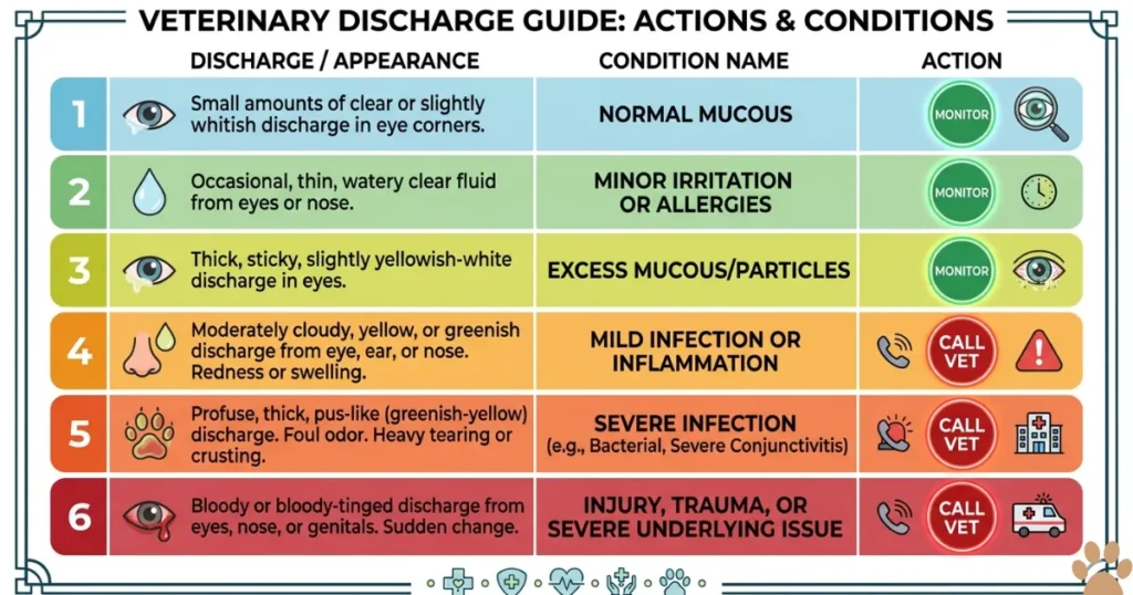
When to Call the Vet: Urgent vs. Monitor Table.
| CALL VET IMMEDIATELY 🔴 | MONITOR AT HOME 24 HRS 🟡 |
| Bulging or asymmetric eye | Clear watery discharge, both eyes open |
| Yellow-green discharge, bilateral and sudden | Mild single-eye tearing after outdoor exposure |
| Bloody or dark brown discharge | Light white crust at inner corner, no squinting |
| Sustained squinting or pawing at the eye | Mild conjunctival redness, eating and drinking normally |
| Visible cysts or dark spots at pupil margin | Blue-grey lens haze without any vision changes |
| Any visible foreign object embedded in eye | — |
If monitoring and any URGENT sign appear within 24 hours, stop monitoring. Call immediately.
EXPERT INSIGHT CALLOUT.
“The picture comparison problem is real. Owners search for dog eye infection pictures and match their Golden to whatever looks closest. In my experience, that leads to KCS being treated as bacterial conjunctivitis repeatedly while the cornea quietly scars. I’d rather an owner describe the texture and color of what they see than try to match a photograph. “Grey-white and ropy versus yellow-green and crusting—those two descriptions change everything about what I prescribe.”
What dog eye health issues should owners watch for?
Dog eye health issues to watch for include discharge color changes from clear to yellow-green, iris darkening, lens opacity, sustained squinting, pawing at the eye, and eye asymmetry. In golden retrievers, ropy grey-white discharge and iris browning are breed-specific warning signs requiring prompt evaluation.
How do I identify dog eye health issues at home?
Assess discharge color and texture, check whether both eyes are equally open, test the menace response by moving your hand slowly toward the eye from the side, and compare iris color to recent photos. Use the GRI Visual Symptom Classifier to map what you see to a probable condition before calling your vet.
What do dog eye infections look like in pictures?
Dog eye infections typically show yellow-green discharge crusting the eyelid, conjunctival redness, and sometimes eyelid swelling. In pictures, bacterial conjunctivitis appears as a paste-like colored material at the eye corners. Ropy grey-white discharge that looks similar is more likely KCS in Golden Retrievers, not infection.
How do dog eye problems look in pictures vs. real life?
Dog eye problems often appear more dramatic in pictures than in early real-life presentations. A cataract shows as a white lens in photos but may appear only as a slight haze to the naked eye initially. KCS discharge looks like standard sleep crust casually but has a distinctive ropy, stringy texture on closer inspection.
What are the common eye ailments in dogs by appearance?
Common eye ailments in dogs, visible by sign, include red conjunctiva for conjunctivitis, hazy lens for cataracts or nuclear sclerosis, ropy discharge for KCS, yellow-green paste for bacterial infection, bulging eyes for canine glaucoma, and iris darkening for uveitis. Each appearance maps to a different condition requiring different treatment.
What is the best eye care routine for dogs?
Wipe clear discharge with a damp, clean cloth daily. Check iris color and lens clarity monthly using a flashlight. For Golden Retrievers over age 7, schedule annual OFA eye exams, including intraocular pressure measurement. Don’t use human eye drops, as their osmolarity and pH differ from canine-formulated products.
Can a dog eye infection spread to humans?
Most canine bacterial conjunctivitis does not spread to humans. Some bacterial causes carry zoonotic potential. Wash your hands after handling a dog with active eye discharge. The AVMA recommends standard hygiene precautions when treating dogs with active ocular infections.
What does normal dog eye discharge look like?
Normal dog eye discharge is a small amount of clear or lightly white material at the inner corner, present after sleep. It wipes away easily with no stringiness. Anything yellow, green, ropy, bloody, or persistent beyond morning is not normal and warrants assessment.
How do vets diagnose eye problems in dogs?
Vets diagnose dog eye problems using a slit lamp for corneal and lens examination, Schirmer tear testing for KCS, fluorescein dye to identify corneal ulcers, and a tonometer for intraocular pressure measurement. Golden Retrievers with suspected uveitis may also undergo anterior segment imaging to assess cyst burden.
What happens if a dog eye infection goes untreated?
Untreated bacterial conjunctivitis can progress to corneal ulceration. Severe ulcers perforate and cause permanent vision loss. Untreated KCS scars the corneal surface. Untreated canine glaucoma destroys the optic nerve within hours of acute pressure elevation. Early treatment prevents the majority of avoidable blindness cases.
Do Golden Retrievers get more eye infections than other breeds?
Yes. Golden Retrievers develop conjunctivitis and KCS at higher rates than most retrievers, driven by their immune profile and periocular coat anatomy. The GRCA recognizes elevated ocular disease risk as a breed-specific concern, and the Golden Retriever Lifetime Study documents higher heritable ocular condition rates in Goldens than in comparative retriever breeds.
What does pigmentary uveitis look like in a Golden Retriever?
Pigmentary uveitis in Golden Retrievers appears as progressive iris darkening or browning, small cysts visible at the pupil margin, and sometimes mild redness. Most owners first notice it as “His eyes look different lately.” The GRCA recommends annual eye screening for Goldens over age 6, specifically to catch this condition early.
How do Golden Retrievers show signs of dry eye differently from Labradors?
Golden Retrievers with KCS typically produce thick, ropy, grey-white discharge more prominently than Labradors with equivalent tear deficiency, likely due to their periocular anatomy and immune profile. The clinical intervention threshold is the same; the Schirmer tear test is below 15 mm per minute, but the visual presentation differs noticeably between the two breeds.
At what age do Golden Retrievers show signs of progressive retinal atrophy?
Golden Retrievers with PRA typically show first behavioral signs between ages 8 and 12, beginning with night blindness before progressing to daytime vision loss. There are no visible eye appearance changes in early PRA, no discharge, no redness, and no opacity. Night hesitation on stairs or reluctance in dim light is the earliest owner-observable signal, per OFA documentation.
My Golden has green discharge and won’t open one eye—is this an emergency?
Yes. Green discharge paired with an eye held shut indicates severe bacterial conjunctivitis with corneal involvement or a corneal ulcer. Both require the same-day veterinary assessment. Don’t apply any eye drops until your vet has ruled out a corneal ulcer, as some drops are contraindicated if the corneal surface is compromised.
Conclusion.
Dog eye health issues in Golden Retrievers follow visible patterns. Knowing the difference between ropy grey-white discharge (KCS), yellow-green crusting (bacterial infection), iris darkening (pigmentary uveitis), and a bulging fixed eye (acute glaucoma) tells you what to do before your vet picks up the phone. Run the GRI Visual Symptom Classifier first: identify the sign, match it to the condition, and act accordingly.
One rule holds across every presentation. When the sign involves pain behavior, vision changes, or a fixed or bulging eye, call your vet the same day. Every other sign earns a 24-hour monitoring window. No longer.
Has your golden shown an eye change that didn’t fit the typical red-eye pictures you found online? Tell me what you saw—the discharge color, the iris appearance, and whether it appeared suddenly or gradually over weeks. Specific descriptions from Golden owners help others in this community recognize signs they might otherwise dismiss as normal aging.

Dr. Nabeel A.
Dr. Nabeel Akram is a Doctor of Veterinary Medicine with more than five years of hands-on experience in animal health, canine nutrition, and preventive care. He is a registered veterinarian with the Pakistan Veterinary Medical Council (PVMC), the statutory body regulating veterinary practice in Pakistan. As the founder of Golden Retriever Insight, Dr. Akram writes and medically reviews every health, nutrition, and grooming guide published on the site. His clinical interests include canine oncology, epilepsy management, and breed-specific nutrition for large breeds — the core topics this site covers. Every article is checked against current veterinary literature and sources such as the Merck Veterinary Manual, AVMA guidance, and peer-reviewed research.